
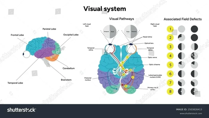
Where is vision processed in the brain? It primarily occurs in the occipital lobe at the back of your head. However, seeing is a complex team effort involving your eyes, optic nerve, and multiple brain regions. This guide breaks down exactly how your brain turns light into the vivid images you experience every day.
Have you ever stopped to marvel at how you can glance across a room and instantly recognize a friend, or how your eyes can track a bird flying through the sky? It feels so effortless, right? But behind the scenes, your brain is performing a massive computational miracle. You might wonder, where is vision processed in the brain? Is it right behind the eyes, or somewhere deeper?
The truth is, vision is not a single event. It is a rapid, synchronized relay race. Light enters your eye, gets converted into electrical signals, and travels through a complex network of brain regions. By the time you actually “see” an image, it has passed through several highly specialized areas. Let us dive into the fascinating neuroscience behind how you see the world.
Key Takeaways
- Primary Processing Location: The main hub for vision is the occipital lobe, located at the back of the brain.
- The Visual Cortex: The primary visual cortex (V1) is the first brain area to receive and organize visual signals from the eyes.
- Two Visual Streams: The brain splits vision into the ventral stream (identifying what things are) and the dorsal stream (locating where things are).
- Beyond the Occipital Lobe: Other brain regions, like the temporal and parietal lobes, help process complex visuals like faces and movement.
- Emotional Connection: How we process visual cues, like facial expressions, deeply impacts our relationships and social interactions.
- Brain Plasticity: If one visual pathway is damaged, the brain can sometimes rewire itself to compensate for the loss.
📑 Table of Contents
- The Journey of Light: From Eye to Brain
- The Occipital Lobe: The Primary Visual Cortex
- The Two Streams of Visual Processing
- How Vision Impacts Our Daily Interactions
- Common Vision Processing Disorders
- Quick Tips for Maintaining Healthy Vision
- Common Mistakes People Make About Vision
- Expert Insights on Visual Neuroscience
- Comparison Table: Visual Pathway Stages
- Conclusion
The Journey of Light: From Eye to Brain
Before we talk about where vision is processed, we need to understand how the signal gets there. Your eyes are not cameras. They are actually direct extensions of your brain. They capture light and translate it into the language of the brain: electricity.
The Role of the Retina
It all starts at the back of your eye, on a thin layer of tissue called the retina. The retina is packed with millions of specialized cells called photoreceptors. There are two main types:
- Rods: These are incredibly sensitive to light and dark. They help you see in low-light conditions, like when you are walking outside at night. However, they do not process color.
- Cones: These require much more light to function. They are responsible for sharp, detailed, and color vision. You have three types of cones, each tuned to red, green, or blue light.
When light hits these photoreceptors, it triggers a chemical reaction that converts the light into an electrical impulse. This is the very first step in visual processing.
The Optic Nerve and Chiasm
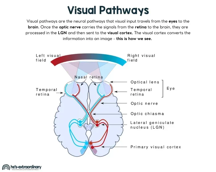
Once the photoreceptors create an electrical signal, it travels through other retinal cells and exits the eye via the optic nerve. Think of the optic nerve as a high-speed fiber-optic cable carrying massive amounts of data.
Here is where it gets interesting. The optic nerves from both eyes meet at a junction called the optic chiasm. At this point, the nerve fibers sort themselves out. The signals from the left visual field of both eyes are routed to the right side of the brain, and the signals from the right visual field are routed to the left side of the brain. This crossover ensures that each hemisphere processes the opposite side of your visual world.
The Occipital Lobe: The Primary Visual Cortex

Visual guide about brain visual processing illustration
Image source: perkins.org
So, where is vision processed in the brain? The primary answer is the occipital lobe. Located at the very back of your skull, this is the brain’s main visual processing center.
When the electrical signals leave the optic chiasm, they travel through the lateral geniculate nucleus (LGN)—a relay station in the thalamus—before arriving at the primary visual cortex, also known as V1 or the striate cortex.
V1 and the Striate Cortex
V1 is the first cortical area to receive visual input. It is incredibly organized. In fact, it contains a precise map of the visual field. If a specific spot on V1 is activated, it corresponds to a specific spot in your visual world.
V1 is responsible for processing basic visual features. It detects:
- Edges and lines: The orientation of objects.
- Spatial frequency: How coarse or fine a pattern is.
- Color contrast: Differences between colors.
- Motion direction: Which way something is moving.
V1 does not “see” a face or a car. It sees a collection of lines, angles, and colors. It breaks the visual world down into its simplest building blocks before sending that information off to other brain areas for complex interpretation.
The Two Streams of Visual Processing
Visual guide about brain visual processing illustration
Image source: hes-extraordinary.com
Once V1 has deconstructed the visual world, the information is sent to other parts of the brain. In the 1980s, neuroscientists discovered that visual processing splits into two distinct pathways, known as the two-stream hypothesis.
The Ventral Stream (“What” Pathway)
The ventral stream travels from the occipital lobe down into the temporal lobe. This pathway is all about object recognition. It answers the question: “What am I looking at?”
This is how you can look at a four-legged animal and instantly know it is a dog, or look at a piece of fruit and know it is an apple. The ventral stream processes details like shape, color, and texture to identify objects. It also plays a massive role in facial recognition. The fusiform face area, located in the temporal lobe, is specifically dedicated to helping you recognize the faces of people you know.
The Dorsal Stream (“Where” Pathway)
The dorsal stream travels from the occipital lobe up into the parietal lobe. This pathway is all about spatial awareness and action. It answers the question: “Where is it, and how do I interact with it?”
If you reach out to grab a coffee cup, your dorsal stream is at work. It processes the location of the cup, its distance, and its orientation, allowing your hand to move accurately to grasp it. It also processes motion, helping you navigate through a crowded room without bumping into people.
How Vision Impacts Our Daily Interactions

Visual guide about brain visual processing illustration
Image source: thumbs.dreamstime.com
Understanding where vision is processed in the brain is not just an academic exercise. It has profound implications for how we interact with others. A huge portion of our visual processing is dedicated to reading people.
When you look at someone, your brain is scanning their face for micro-expressions. It is reading their body language. This visual data is processed incredibly fast, often without you even realizing it.
For instance, when someone you care about shuts down during a conversation, their face might go blank. Your brain immediately registers this visual shift. Recognizing these subtle visual cues is vital for healthy relationships. In fact, stonewalling is the most overlooked form of emotional abuse precisely because the visual cue of a blank face can be easily ignored or misinterpreted by the observer’s brain.
Visual Cues in Attraction and Connection
Have you ever wondered why you are instantly drawn to someone? Much of it comes down to visual processing. Your ventral stream is rapidly assessing facial symmetry and familiarity. Meanwhile, your brain is releasing chemicals based on what you see.
Understanding how the brain processes attraction can be eye-opening. It helps us realize that what we perceive as “chemistry” is actually a complex neurological event. If you want to understand the deeper implications of how we view others visually, it is worth exploring the difference between attraction and sexual objectification. Healthy visual processing allows us to see a whole person, while objectification reduces a person to a collection of visual parts.
How Trauma Alters Visual Processing
Our past experiences physically shape how our brains process visual information. If someone has experienced trauma, their visual processing can become hyper-vigilant. Their brain might be wired to scan faces for anger or threat, bypassing the normal, relaxed visual processing.
This is why a person who uses humor as a defense mechanism might still struggle with connection. You might wonder, what are the odds a funny person is traumatized? The answer is quite high, and their altered visual processing often reflects this underlying pain. Their brain is working overtime to visually assess safety, making it hard to relax and truly “see” the people around them.
Common Vision Processing Disorders
Because visual processing involves so many different brain regions, damage to specific areas can cause very specific and strange visual deficits.
Visual Agnosia
If the ventral stream is damaged, a person might develop visual agnosia. They can see perfectly fine—their eyes work, and V1 processes the light—but they cannot recognize what they are looking at. A person with visual agnosia might look at a key and describe it as a “small metal object,” but they cannot name it. If they are allowed to feel it, they instantly know what it is.
Blindsight
Blindsight is perhaps the most baffling visual disorder. It occurs when V1 is damaged, causing the person to be clinically blind in a specific area of their visual field. However, if you throw a ball at them, they might catch it. They cannot consciously see the ball, but their dorsal stream (the “where” pathway) is still processing the spatial information. The brain knows where the object is, even if the conscious mind does not.
Akinetopsia
This is a rare condition caused by damage to area V5 in the dorsal stream. People with akinetopsia cannot perceive motion. They see the world as a series of static frames. For example, if they watch a car drive by, they would see the car in one spot, and then suddenly in another spot, with no sense of movement in between. Pouring a cup of tea becomes a dangerous task because they cannot see the liquid rising.
Quick Tips for Maintaining Healthy Vision
Your brain’s visual processing system relies entirely on the quality of the data it receives from your eyes. If your eyes are unhealthy, your brain suffers. Here are some quick tips to keep your visual system sharp:
- Eat for your eyes: Consume foods rich in Vitamin A, lutein, and omega-3 fatty acids. Think carrots, leafy greens, and salmon.
- Wear sunglasses: Protect your retinas from harmful UV rays to prevent long-term photoreceptor damage.
- Take screen breaks: Follow the 20-20-20 rule. Every 20 minutes, look at something 20 feet away for 20 seconds to reduce eye strain.
- Get regular eye exams: Catching issues early ensures your brain gets the clearest visual input possible.
Common Mistakes People Make About Vision
There are a lot of myths about how we see. Here are the most common mistakes people make about visual processing:
- Myth 1: Vision happens in the eyes. Reality: The eyes only capture light. Vision happens in the brain. Without the brain to interpret the signals, the eyes are useless.
- Myth 2: We see everything in perfect detail. Reality: Your peripheral vision is incredibly blurry and mostly black-and-white. Your brain fills in the gaps and makes you think you have a sharp, colorful picture of the whole world.
- Myth 3: The brain processes vision like a camera recording. Reality: The brain actively constructs your visual reality. It takes shortcuts, fills in blind spots, and heavily filters information based on what it deems important.
Expert Insights on Visual Neuroscience
Neuroscientists are constantly discovering new layers of complexity in visual processing. One of the most exciting areas of research is predictive processing. Experts now believe that your brain is not just passively receiving visual data; it is actively predicting what it expects to see next.
Your brain uses past experiences to generate a model of the world. When visual data comes in, the brain compares it to the prediction. If the prediction is correct, it ignores the details. If there is an error, it updates the model. This is why you can read a paragraph full of typos—your brain predicts the words and does not bother processing every single letter. This predictive nature also explains why optical illusions work; they trick your brain’s predictions.
Comparison Table: Visual Pathway Stages
To help you visualize the visual processing journey, here is a breakdown of the stages:
| Stage | Brain Region | Primary Function |
|---|---|---|
| 1. Capture | Retina (Rods and Cones) | Converts light into electrical signals |
| 2. Relay | Optic Nerve and Thalamus (LGN) | Transmits and sorts visual signals to the cortex |
| 3. Basic Processing | Primary Visual Cortex (V1) | Detects edges, lines, colors, and motion |
| 4. Object Recognition | Ventral Stream (Temporal Lobe) | Identifies “what” objects and faces are |
| 5. Spatial Action | Dorsal Stream (Parietal Lobe) | Identifies “where” objects are and guides movement |
Conclusion
So, where is vision processed in the brain? While the occipital lobe serves as the primary hub, vision is truly a whole-brain experience. From the moment light hits your retina to the instant you recognize a loved one’s face, your brain is working at lightning speed. It deconstructs light, routes it through the thalamus, processes basic features in V1, and then splits the data into the ventral and dorsal streams to give meaning and spatial context to the world.
Next time you gaze out a window or look into someone’s eyes, take a moment to appreciate the incredible neural symphony playing out inside your skull. Understanding how your brain processes vision not only deepens your appreciation for the miracle of sight, but it can also make you more mindful of how you visually engage with the people around you. When we truly “see” others—reading their expressions, respecting their boundaries, and acknowledging their presence—we build deeper, more meaningful connections.
Frequently Asked Questions
What part of the brain is responsible for vision?
The occipital lobe, located at the back of the brain, is the primary center for visual processing. It houses the primary visual cortex, which receives and interprets signals from the eyes.
What happens if the occipital lobe is damaged?
Damage to the occipital lobe can cause vision loss, blind spots, or difficulty recognizing objects and faces. In rare cases, it can cause blindsight, where a person cannot consciously see but can still respond to visual stimuli.
How does the brain process visual information?
The brain processes visual information in stages. First, the retina converts light into electrical signals. These signals travel to the occipital lobe for basic processing, then split into two streams: one for identifying objects and one for locating them in space.
Can the brain adapt if vision is lost?
Yes, the brain has a remarkable ability called neuroplasticity. If vision is lost, the brain can sometimes rewire itself. For example, the visual cortex might start processing touch or sound, which is why blind individuals often have heightened hearing or tactile senses.
What is the difference between the ventral and dorsal streams?
The ventral stream travels to the temporal lobe and is responsible for object recognition (the “what”). The dorsal stream travels to the parietal lobe and handles spatial awareness and movement (the “where”).
Why do optical illusions trick the brain?
Optical illusions exploit the brain’s predictive processing. Your brain takes shortcuts based on past experiences to guess what you are seeing. Illusions provide misleading visual data that causes the brain to make an incorrect prediction.
